Safer medical imaging with microwaves
The goal of the research is to develop an alternative method to x-ray imaging that is safer and uses nothing stronger than radio frequency waves.
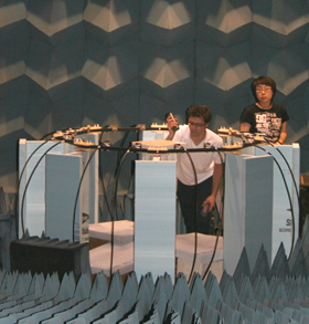
Research being conducted by Prof. Mahta Moghaddam and her group could lead to safer medical imaging practices in hospitals and labs. The goal is to develop an alternative and complementary method to x-ray imaging that is safer and uses nothing stronger than the radio frequency waves present when operating a cell phone. A recent experiment, described in the video below, proved the success of a preliminary stage in the development of this new technology.
Background
Early detection of breast cancer provides perhaps the best chances for long-term survival rate, resulting in a better than 95% chance of survival for five years or longer. The current gold standard for breast imaging is with x-ray mammography; however, it has numerous risks and pitfalls. These include the lack of patient safety due to the ionizing radiation, patient discomfort, and especially the inherent imaging ambiguities that regularly result in a large percentage of false positives (which lead to unnecessary biopsies) and false negatives (which miss malignant tumors).
Therefore, researchers are actively looking for a better way to find tumors in the body.
Recent advances in microwave imaging show promise for improved detection and diagnosis of small early-stage tumors, due to the distinct electrical contrast of the cancerous tumors with respect to healthy breast tissue. Microwave breast imaging uses low-power radio frequency signals, similar to cell phones. The main challenge in microwave imaging is in converting the rich information content of its measurements to quantitative tissue properties to allow specific diagnostics. This process is called inverse scattering.
Research details
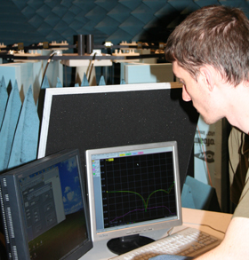
While microwave inverse scattering has been extensively studied in theory, successful demonstrations are few and far-between. Inverse scattering algorithms estimate material contrasts by comparing numerical predictions of microwave scattering models to measurements, and progressively tune the models (using nonlinear optimization techniques) to achieve a good match with measurements. The scattering model is typically a complicated integral equation that calculates the complex-valued electric fields. However, the same scattered field cannot be measured directly in experiment; only a voltage response at the output of an antenna can be measured.
One of the most difficult aspects of experimental inverse scattering is relating the idealized electric field predictions in the algorithms to actual antenna voltage measurements in experiment. For a long time this has been an outstanding problem for validating inversion algorithms, with partial solutions typically in the form of applying empirical calibration factors to voltage measurements to convert them to complex field quantities or scattering cross sections. Such techniques only partially solve the calibration problem since in reality these factors are not simple constants and can vary significantly with observation angle, frequency, material properties, etc.

 Enlarge

 Enlarge
Results and future direction
Prof. Mahta Moghaddam and her students have recently developed a new technique to forge the missing link between the complex electric fields predicted by theoretical models and voltage ratio measurements typically available in experiments, and, for the first time, demonstrated a microwave inverse scattering technique with end-to-end, self-contained absolute calibration. The group is now developing the technique further to achieve a clinically representative microwave imaging system.
 MENU
MENU 
